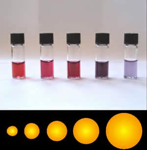
Colloidal gold is a suspension (or colloid) of submicrometre-size particles of gold in a fluid, usually water. The liquid is usually either an intense red colour (for particles less than 100Â nm) or blue/purple (for larger particles). Due to the unique optical, electronic, and molecular-recognition properties of gold nanoparticles, they are the subject of substantial research, with applications in a wide variety of areas, including electron microscopy, electronics, nanotechnology, and materials science.
Properties and applications of colloidal gold nanoparticles strongly depend upon their size and shape. For example, rodlike particles have both transverse and longitudinal absorption peak, and anisotropy of the shape affects their self-assembly.
History

Known, or at least used (perhaps proceeding by accident without much understanding of the process) since ancient times, the synthesis of colloidal gold was crucial to the 4th-century Lycurgus Cup, which changes colour depending on the direction of the light. Later it was used as a method of staining glass.
A so-called Elixir of Life, a potion made from gold, was discussed, if not actually manufactured, in ancient times. In the 16th century, the alchemist Paracelsus claimed to have created a potion called Aurum Potabile (Latin: potable gold). In The newe iewell of health, 1576, translator George Baker promoted the use and preparation of potable gold, along with other "most excellent secretes of phisicke and philosophie".
In the 17th century, the glass-colouring process was refined by Andreus Cassius and Johann Kunckel, allowing them to produce a striking deep-ruby colored form of glass. In 1842, John Herschel invented a photographic process called chrysotype (from the Greek χÏῡσός meaning "gold") that used colloidal gold to record images on paper.
Modern scientific evaluation of colloidal gold did not begin until Michael Faraday's work of the 1850s. Paracelsus' work is known to have inspired Faraday to prepare the first pure sample of colloidal gold, which he called 'activated gold', in 1857. He used phosphorus to reduce a solution of gold chloride.
For a long time, the composition of the Cassius ruby-gold was unclear. Several chemists suspected it to be a gold tin compound, due to its preparation. Faraday was the first to recognize that the color was actually due to the minute size of the gold particles.
In 1898, Richard Adolf Zsigmondy prepared the first colloidal gold in diluted solution. Apart from Zsigmondy, Theodor Svedberg, who invented ultracentrifugation, and Gustav Mie, who provided the theory for scattering and absorption by spherical particles, were also interested in understanding synthesis and properties of colloidal gold.
Synthesis

Generally, gold nanoparticles are produced in a liquid ("liquid chemical methods") by reduction of chloroauric acid (H[AuCl4]). After dissolving H[AuCl4], the solution is rapidly stirred while a reducing agent is added. This causes Au3+ ions to be reduced to neutral gold atoms. As more and more of these gold atoms form, the solution becomes supersaturated, and gold gradually starts to precipitate in the form of sub-nanometer particles. The rest of the gold atoms that form stick to the existing particles, and, if the solution is stirred vigorously enough, the particles will be fairly uniform in size.
To prevent the particles from aggregating, some sort of stabilizing agent that sticks to the nanoparticle surface is usually added. Also, gold colloids can be synthesised without stabilizers by laser ablation in liquids.
They can be functionalized with various organic ligands to create organic-inorganic hybrids with advanced functionality.
Turkevich method
The method pioneered by J. Turkevich et al. in 1951 and refined by G. Frens in the 1970s, is the simplest one available. In general, it is used to produce modestly monodisperse spherical gold nanoparticles suspended in water of around 10â€"20 nm in diameter. Larger particles can be produced, but this comes at the cost of monodispersity and shape. It involves the reaction of small amounts of hot chloroauric acid with small amounts of sodium citrate solution. The colloidal gold will form because the citrate ions act as both a reducing agent and a capping agent.
Recently, the evolution of the spherical gold nanoparticles in the Turkevich reaction has been elucidated. It is interesting to note that extensive networks of gold nanowires are formed as a transient intermediate. These gold nanowires are responsible for the dark appearance of the reaction solution before it turns ruby-red.
To produce larger particles, less sodium citrate should be added (possibly down to 0.05%, after which there simply would not be enough to reduce all the gold). The reduction in the amount of sodium citrate will reduce the amount of the citrate ions available for stabilizing the particles, and this will cause the small particles to aggregate into bigger ones (until the total surface area of all particles becomes small enough to be covered by the existing citrate ions).
Brust method
This method was discovered by Brust and Schiffrin in the early 1990s, and can be used to produce gold nanoparticles in organic liquids that are normally not miscible with water (like toluene). It involves the reaction of a chlorauric acid solution with tetraoctylammonium bromide (TOAB) solution in toluene and sodium borohydride as an anti-coagulant and a reducing agent, respectively.
Here, the gold nanoparticles will be around 5â€"6 nm. NaBH4 is the reducing agent, and TOAB is both the phase transfer catalyst and the stabilizing agent.
It is important to note that TOAB does not bind to the gold nanoparticles particularly strongly, so the solution will aggregate gradually over the course of approximately two weeks. To prevent this, one can add a stronger binding agent, like a thiol (in particular, alkanethiols), which will bind to gold, producing a near-permanent solution. Alkanethiol protected gold nanoparticles can be precipitated and then redissolved. Some of the phase transfer agent may remain bound to the purified nanoparticles, this may affect physical properties such as solubility. In order to remove as much of this agent as possible, the nanoparticles must be further purified by soxhlet extraction.
Perrault method
This approach, discovered by Perrault and Chan in 2009, uses hydroquinone to reduce HAuCl4 in an aqueous solution that contains 15 nm gold nanoparticle seeds. This seed-based method of synthesis is similar to that used in photographic film development, in which silver grains within the film grow through addition of reduced silver onto their surface. Likewise, gold nanoparticles can act in conjunction with hydroquinone to catalyze reduction of ionic gold onto their surface. The presence of a stabilizer such as citrate results in controlled deposition of gold atoms onto the particles, and growth. Typically, the nanoparticle seeds are produced using the citrate method. The hydroquinone method complements that of Frens, as it extends the range of monodispersed spherical particle sizes that can be produced. Whereas the Frens method is ideal for particles of 12-20 nm, the hydroquinone method can produce particles of at least 30â€"300 nm.
Martin method
This simple method, discovered by Martin and Eah in 2010, generates nearly monodisperse “naked†gold nanoparticles in water. Precisely controlling the reduction stoichiometry by simply adjusting the ratio of NaBH4-NaOH ions to HAuCl4-HCl ions within the “sweet zone,†along with heating, enables reproducible diameter tuning between 3-6 nm. The aqueous particles are colloidally stable due to their high charge from the excess ions in solution. These particles can be coated with various hydrophilic functionalities, or mixed with hydrophobic molecules for applications in non-polar solvents. Interestingly, in non-polar solvents the nanoparticles curiously remain highly charged, and self-assemble on liquid droplets to form 2D monolayer films of monodisperse nanoparticles.
Nanotech applications
Bacillus licheniformis can be used in synthesis of gold nanocubes. Researchers have synthesized gold nanoparticles with sizes between 10 to 100 nanometres. Gold nanoparticles are usually synthesized at high temperatures, in organic solvents and using toxic reagents. The bacteria produce them in much milder conditions.
As pointed out before, the precise size control with a low polydispersity of spherical gold nanoparticles remains difficult for particles larger than 30 nm. In order to provide maximum control on the NP structure along with synthetic straightforwardness, Navarro and co-workers used a ‘one-pot’ protocol, relying on a modified Turkevitch-Frens procedure. The method consist in the addition of sodium acetylacetonate (Na(acac) allowing the complexation and reduction of AuIII to AuI) immediately followed by the addition of sodium citrate (reduction of AuI to Au0). For this experiment, sodium acetylacetonate purity is really important. The concentration of sodium acetylacetonate directly impacts the nuclei numbers inducing a size evolution of the gold core up to 90 nm with a narrow size distribution. Practically, the tetrachloroaurate and sodium citrate concentration are fixed to 0.30 mM and 0.255 mM (ratio sodium citrate to gold: 0.85). In these conditions, and without sodium acetylacetonate, the obtained nanoparticles precipitate immediately. The concentration of sodium acetylacetonate can be tuned from 0.33 mM to 1.0 mM. The addition of the Na(acac) and sodium citrate to the yellow boiling gold ions solution results in a colorless one indicating the complexation and reduction of AuIII to AuI. Within a few minutes, the solution turns light blue-purple, featuring the formation of the gold nuclei. The small crystalline structures will then diffuse over the solution allowing the growth of the final spherical particles.
Sonolysis
Another method for the experimental generation of gold particles is by sonolysis. The first method of this type was invented by Baigent and Müller. This work pioneered the use of ultrasound to provide the energy for the processes involved and allowed the creation of gold particles with a diameter of under 10 nm. In another method using ultrasound, the reaction of an aqueous solution of HAuCl4 with glucose, the reducing agents are hydroxyl radicals and sugar pyrolysis radicals (forming at the interfacial region between the collapsing cavities and the bulk water) and the morphology obtained is that of nanoribbons with width 30â€"50 nm and length of several micrometers. These ribbons are very flexible and can bend with angles larger than 90°. When glucose is replaced by cyclodextrin (a glucose oligomer), only spherical gold particles are obtained, suggesting that glucose is essential in directing the morphology toward a ribbon.
Block copolymer-mediated method
An economical, environmentally benign and fast synthesis methodology for gold nanoparticles using block copolymer has been developed by Sakai et al. In this synthesis methodology, block copolymer plays the dual role of a reducing agent as well as a stabilizing agent. The formation of gold nanoparticles comprises three main steps: reduction of gold salt ion by block copolymers in the solution and formation of gold clusters, adsorption of block copolymers on gold clusters and further reduction of gold salt ions on the surfaces of these gold clusters for the growth of gold particles in steps, and finally its stabilization by block copolymers. But this method usually has a limited-yield (nanoparticle concentration), which does not increase with the increase in the gold salt concentration. Recently, Ray et al. demonstrated that the presence of an additional reductant (trisodium citrate) in 1:1 molar ratio with gold salt enhances the yield by manyfold at ambient conditions and room temperature.
“Green Chemistry†based methods
Phytochemicals found in various plant sources have been utilized as a means of developing a more economical and environmentally friendly synthetic pathway in the formation of gold nanoparticles. In accordance to the principles of “green chemistry,†these methods employ the use of nontoxic chemicals, marginal energy consumption, renewable materials, and environmentally benign solvents to minimize the use, disposal, and health repercussions of hazardous chemicals. Additionally, these methods provide a more efficient synthetic pathway through a one-step process without the use of supplementary surfactants or polymers, capping agents, or templates to restrict agglomeration of the gold nanoparticles. This method is effective in producing well-defined gold nanoparticles since the phytochemicals perform a dual role as both a reducing agent of gold and as a stabilizer in the formation of a sturdy coating on the nanoparticles. One “green†method that has been employed in the formation of gold nanoparticles utilizes the phytochemicals and polyphenols in Darjeeling black tea leaves, with water acting as a benign solvent at room temperature. The phytochemicals in the black tea reduce HAuCl4 to its salt and stabilize the aggregation of the gold atoms as the nanoparticle is formed. In addition to the “green†benefits of using black tea, the size of the nanoparticle is influenced by the concentration of the tea, and the absorbance and size of the gold nanoparticles formed can be easily determined using UV-Vis spectrometry abiding that an increase in λmax correlates to an increase in the size of the nanoparticle. Other “green†methods that have been studied and employed include the use of Elettaria cardamomum (cardamom) and cinnamon in the synthesis of gold nanoparticles, as well as Syzygium aromaticum (cloves) in the formation of copper nanoparticles and table sugar in the formation of silver nanoparticles.
Electron microscopy
Colloidal gold and various derivatives have long been among the most widely used labels for antigens in biological electron microscopy. Colloidal gold particles can be attached to many traditional biological probes such as antibodies, lectins, superantigens, glycans, nucleic acids, and receptors. Particles of different sizes are easily distinguishable in electron micrographs, allowing simultaneous multiple-labelling experiments.
In addition to biological probes, gold nanoparticles can be transferred to various mineral substrates, such as mica, single crystal silicon, and atomically flat gold(III), to be observed under atomic force microscopy (AFM).
Medical research
Drug delivery system
Gold nanoparticles can be used to optimize the biodistribution of drugs to diseased organs, tissues or cells, in order to improve and target drug delivery. It is important to realize that the nanoparticle-mediated drug delivery is feasible only if the drug distribution is otherwise inadequate. These cases include drug targeting of difficult, unstable molecules (proteins, siRNA, DNA), delivery to the difficult sites (brain, retina, tumors, intracellular organelles) and drugs with serious side effects (e.g. anti-cancer agents). The performance of the nanoparticles depends on the size and surface functionalities in the particles. Also, the drug release and particle disintegration can vary depending on the system (e.g. biodegradable polymers sensitive to pH). An optimal nanodrug delivery system ensures that the active drug is available at the site of action for the correct time and duration, and their concentration should be above the minimal effective concentration (MEC) and below the minimal toxic concentration (MTC).
Gold nanoparticles are being investigated as carriers for drugs such as Paclitaxel. The administration of hydrophobic drugs require molecular encapsulation and it is found that nanosized particles are particularly efficient in evading the reticuloendothelial system.
Gold nanoparticles are also used to circumvent multidrug resistance (MDR) mechanisms. Mechanisms of MDR include decreased uptake of drugs, reduced intracellular drug concentration by activation of the efflux transporters, modifications in cellular pathways by altering cell cycle checkpoints, increased metabolism of drugs, induced emergency response genes to impair apoptotic pathways and altered DNA repair mechanisms.
Tumor detection
In cancer research, colloidal gold can be used to target tumors and provide detection using SERS (Surface Enhanced Raman Spectroscopy) in vivo. These gold nanoparticles are surrounded with Raman reporters, which provide light emission that is over 200 times brighter than quantum dots. It was found that the Raman reporters were stabilized when the nanoparticles were encapsulated with a thiol-modified polyethylene glycol coat. This allows for compatibility and circulation in vivo. To specifically target tumor cells, the pegylated gold particles are conjugated with an antibody (or an antibody fragment such as scFv), against, e.g. Epidermal growth factor receptor, which is sometimes overexpressed in cells of certain cancer types. Using SERS, these pegylated gold nanoparticles can then detect the location of the tumor.
Gold nanoparticles accumulate in tumors, due to the leakiness of tumor vasculature, and can be used as contrast agents for enhanced imaging in a time-resolved optical tomography system using short-pulse lasers for skin cancer detection in mouse model. It is found that intravenously administrated spherical gold nanoparticles broadened the temporal profile of reflected optical signals and enhanced the contrast between surrounding normal tissue and tumors.
Therefore, gold nanoparticles have the potential to join numerous therapeutic functions into a single platform, by targeting specific tumor cells, tissues and organs. Actually, Conde et al. reported the evaluation of the inflammatory response and therapeutic siRNA silencing via RGD-nanoparticles in a lung cancer mouse model. This study reported the use of siRNA/RGD gold nanoparticles capable of targeting tumor cells in two lung cancer xenograft mouse models, resulting in successful and significant c-Myc oncogene downregulation followed by tumor growth inhibition and prolonged survival of the animals. This delivery system can achieve translocation of siRNA duplexes directly into the tumour cell cytoplasm and accomplish successful silencing of an oncogene expression. Actually, RGD/siRNA-AuNPs can target preferentially and be taken up by tumor cells via integrin αvβ3-receptor-mediated endocytosis with no cytotoxicity, showing that can accumulate in tumor tissues overexpressing αvβ3 integrins and selectively delivered c-Myc siRNA to suppress tumor growth and angiogenesis.
Gene therapy
Gene therapy is receiving increasing attention and, in particular, small-interference RNA (siRNA) shows importance in novel molecular approaches in the knockdown of specific gene expression in cancerous cells. The major obstacle to clinical application is the uncertainty about how to deliver therapeutic siRNAs with maximal therapeutic impact. Gold nanoparticles have shown potential as intracellular delivery vehicles for siRNA oligonucleotides with maximal therapeutic impact.
Recently, Conde et al. provided evidence of in vitro and in vivo RNAi triggering via the synthesis of a library of novel multifunctional gold nanoparticles, using a hierarchical approach including three biological systems of increasing complexity: in vitro cultured human cells, in vivo freshwater polyp (Hydra vulgaris), and in vivo mice models. The authors developed effective conjugation strategies to combine, in a highly controlled way, specific biomolecules to the surface of gold nanoparticles such as: (a) biofunctional spacers: Poly(ethylene glycol) (PEG) spacers used to increase solubility and biocompatibility; (b) cell penetrating peptides such as TAT and RGD peptides: A novel class of membrane translocating agents named cell penetrating peptides (CPPs) that exploit more than one mechanism of endocytosis to overcome the lipophilic barrier of the cellular membranes and deliver large molecules and even small particles inside the cell for their biological actions; and (c) siRNA complementary to a master regulator gene, the protooncogene c-myc, were bond covalently (thiol-siRNA) and ionically (naked/unmodified siRNA) to gold nanoparticles.
Gold nanoparticles have also shown potential as intracellular delivery vehicles for antisense oligonucleotides (ssDNA,dsDNA) by providing protection against intracellular nucleases and ease of functionalization for selective targeting. Recently, Conde et al. developed a new theranostic system capable of intersecting all RNA pathways: from gene specific downregulation to silencing the silencers, i.e. siRNA and miRNA pathways. The authors reported the development gold nanoparticles functionalized with a fluorophore labeled hairpin-DNA, i.e. gold nanobeacons, capable of efficiently silencing single gene expression, exogenous siRNA and endogenous miRNAs while yielding a quantifiable fluorescence signal directly proportional to the level of silencing. This method describes a gold nanoparticle-based nanobeacon as an innovative theranostic approach for detection and inhibition of sequence-specific DNA and RNA for in vitro and ex vivo applications. Under hairpin configuration, proximity to gold nanoparticles leads to fluorescence quenching; hybridization to a complementary target restores fluorescence emission due to the gold nanobeacons’ conformational reorganization that causes the fluorophore and the gold nanoparticle to part from each other. This concept can easily be extended and adapted to assist the in vitro evaluation of silencing potential of a given sequence to be later used for ex vivo gene silencing and RNAi approaches, with the ability to monitor real-time gene delivery action.
Photothermal agents
Gold nanorods are being investigated as photothermal agents for in-vivo applications. Gold nanorods are rod-shaped gold nanoparticles whose aspect ratios tune the surface plasmon resonance (SPR) band from the visible to near-infrared wavelength. The total extinction of light at the SPR is made up of both absorption and scattering. For the smaller axial diameter nanorods (~10Â nm), absorption dominates, whereas for the larger axial diameter nanorods (>35Â nm) scattering can dominate. As a consequence, for in-vivo applications, small diameter gold nanorods are being used as photothermal converters of near-infrared light due to their high absorption cross-sections. Since near-infrared light transmits readily through human skin and tissue, these nanorods can be used as ablation components for cancer, and other targets. When coated with polymers, gold nanorods have been known to circulate in-vivo for greater than 15 hours half-life. Apart from rod-like gold nanoparticles, also spherical colloidal gold nanoparticles are recently used as markers in combination with photothermal single particle microscopy.
Radiotherapy dose enhancer
Following work by Hainfield et al. there has been considerable interest in the use of gold and other heavy-atom containing nanoparticles to enhance the dose delivered to tumors. Since the gold nanoparticles are taken up by the tumors more than the nearby healthy tissue, the dose is selectively enhanced. The biological effectiveness of this type of therapy seems to be due to the local deposition of the radiation dose near the nanoparticles. This mechanism is the same as occurs in heavy ion therapy.
Detection of toxic gas
Researchers have developed simple inexpensive methods for on-site detection of hydrogen sulfide H
2S present in air based on the antiaggregation of gold nanoparticles (AuNPs). Dissolving H
2S into a weak alkaline buff solution leads to the formation of HS-, which can stabilize AuNPs and ensure they maintain their red color allowing for visual detection of toxic levels of H
2S.
See also
- Colloidal silver
References
Further reading
- Boisselier, E.; Astruc, D (2009). "Gold nanoparticles in nanomedicine: preparations, imaging, diagnostics, therapies and toxicity". Chemical Society Reviews 38 (6). pp. 1759â€"1782. doi:10.1039/b806051g. - " This critical review provides an overall survey of the basic concepts and up-to-date literature results concerning the very promising use of gold nanoparticles (AuNPs) for medicinal applications."
- Conde, J.; Doria, G; Baptista, P (2012). "Noble Metal Nanoparticles Applications in Cancer". Journal of Drug Delivery 2012 (6). pp. 1â€"12. doi:10.1155/2012/751075. - "This review provides insights of the available noble metal nanoparticles for cancer therapy, with particular focus on those already being translated into clinical settings."
- Conde, J.; Rosa, J; Lima, J.C.; Baptista, P.V. (2012). "Nanophotonics for Molecular Diagnostics and Therapy Applications". International Journal Photoenergy 2012.Â
External links
- Moriarty, Philip. "Au â€" Gold Nanoparticle". Sixty Symbols. Brady Haran for the University of Nottingham.Â
- Point-by-point methods for citrate synthesis and hydroquinone synthesis of gold nanoparticles are available here.


0 komentar :
Posting Komentar